| layout | title | show-avatar |
|---|---|---|
page |
Electron Microscopy |
false |
Macrostomum flatworms are transparent which allows us to study they internal organs - in particular their genital morphology - in detail without the need for dissections or histological preparations. However there always remains the lingering suspicion that some important aspects of morphology might be missed because they are not easily visible in the light microscope. To address this issue I am using a specialized electron microscopy technique called Serial Block Face Scanning Electron Microscopy (SBFSEM) to produce serial sections of some animals and examine their morphology in more detail. SBFSEM combines a standard SEM with a microtome that is mounted inside the microscope. The machine scans the block with the electron beam and the backscatter is then analyzed yielding images that can rival TEM quality. After scanning the microtome in the machine removes a slice and then repeats the process. This is nicely visualized in this video from Gatan.
{% include youtubePlayer.html id="PWbLA7WMd34" %}
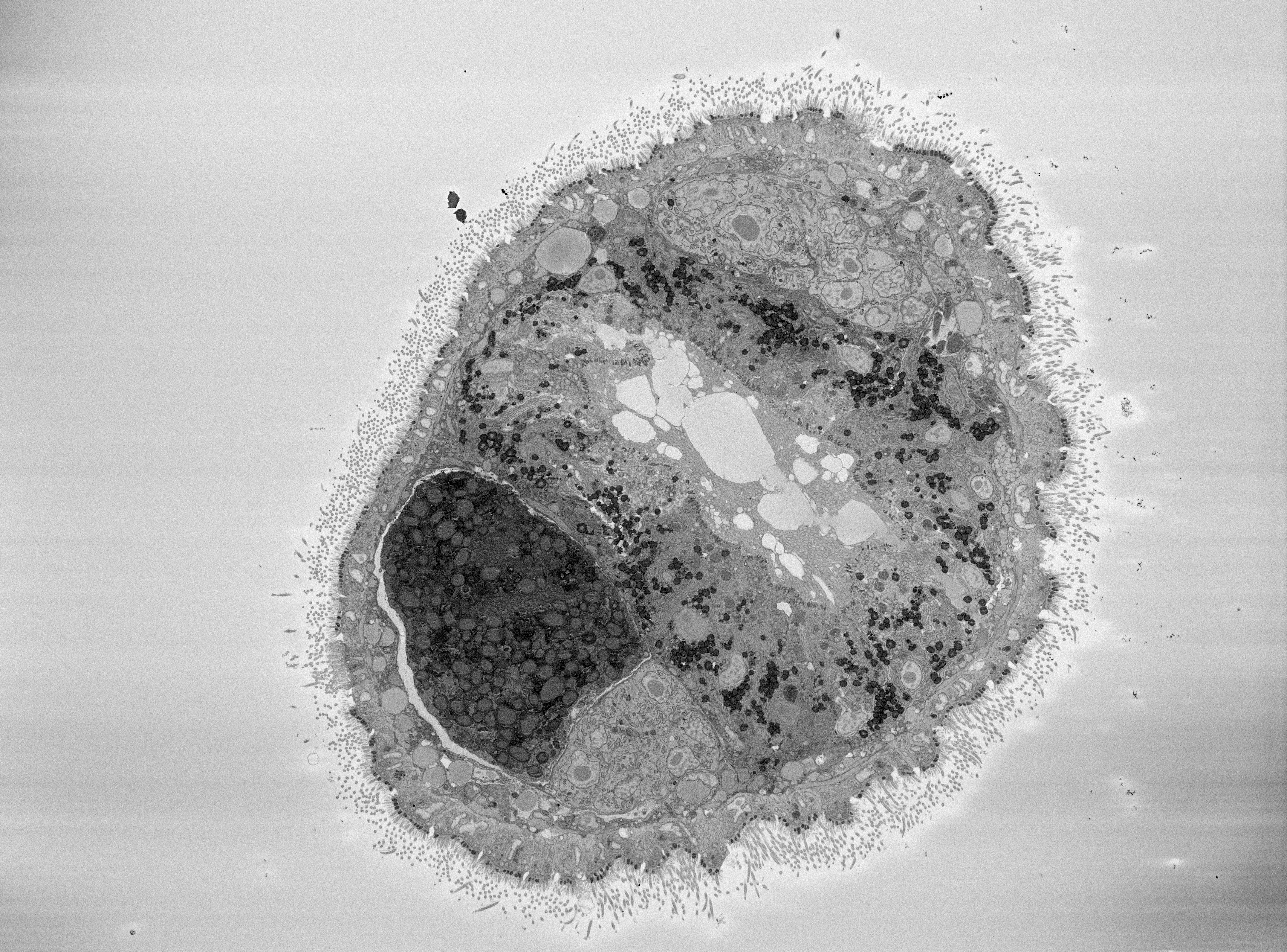
Using this technique we can get images of up to 7nm resolution. In most cases we do not use the maximum resolution because we are interested in the general arrangement of the tissues and not in cell internal processes. Resolution also trades off heavily with scan time and scanning a whole animal at high resolution would easily take two to three weeks! Lower resolutions also give us really nice images:
On this image we see a horizontal section through an entire animal. The thin appendages on the periphery of the animal are cilia that cover the entire surface of the worms body. Smaller cilia are used for gliding locomotion and beat in a synchronized wave. Larger cilia are immobile and used for the detection of sensory stimulus. Below the cilia we see the epidermis cells with very prominent nuclei. These nuclei are not round like in most other cells but multi lobed which sometimes gives them a strange appearance. Centrally we can also see some cilia again but this time they are actually on the inside of the animal lining the gut. Cilia in the gut are longer than the ones on the epidermis and they are less synchronized in their beating. They are used to transport food particles up and down the digestive tract. Something that is quite important for these animals, since they have a one-way digestive system with a mouth opening but no anus (It's not as gross as it sounds). On the left left of the gut we can see a dark heavily stained region. This is a developing oocyte that contains yolk vesicles. Because of the large amounts of membrane present in these vesicles they attract a lot of the osmium used to stain the sample. Right next to the egg we can see the much lighter colored ovary.
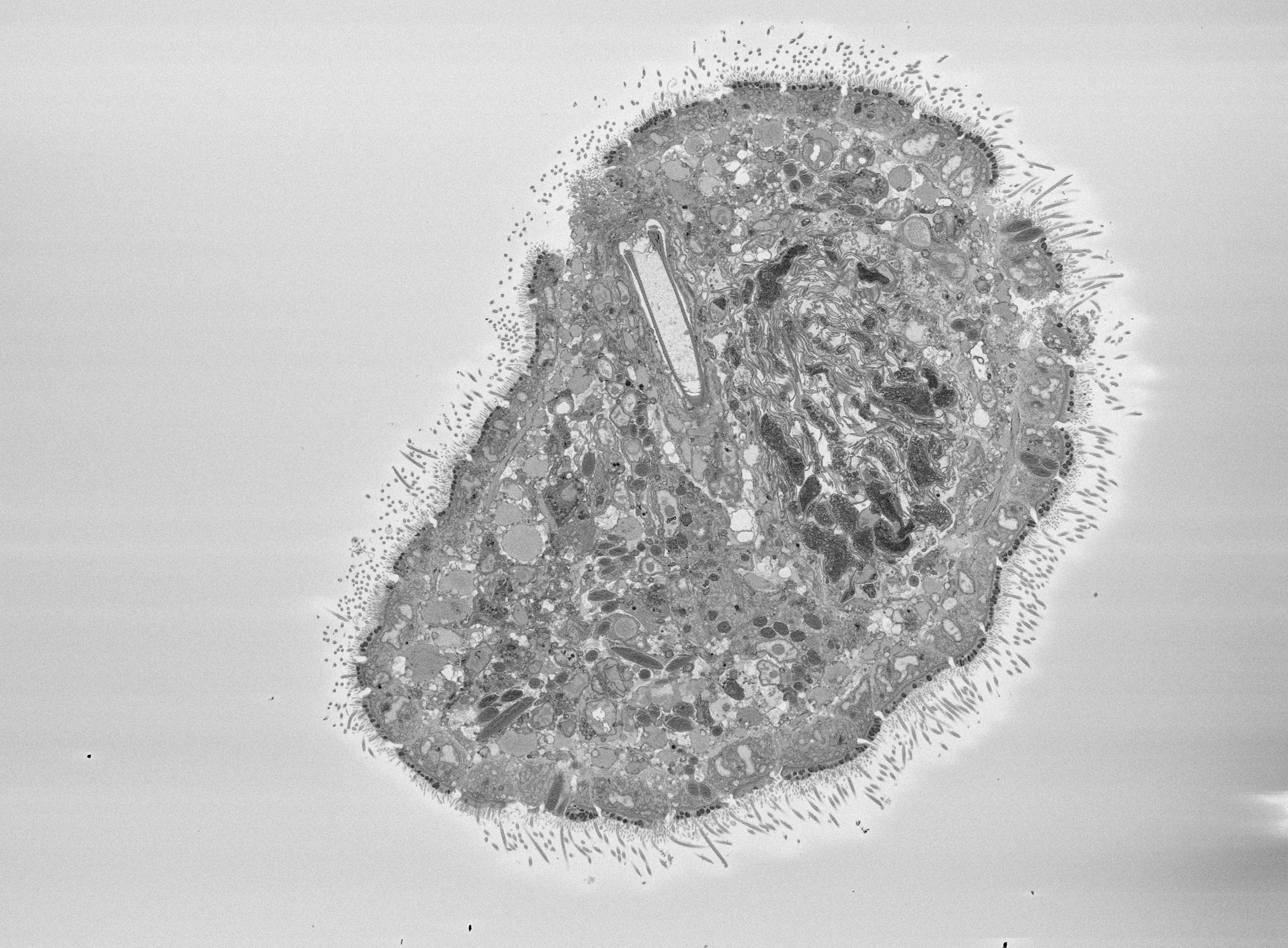
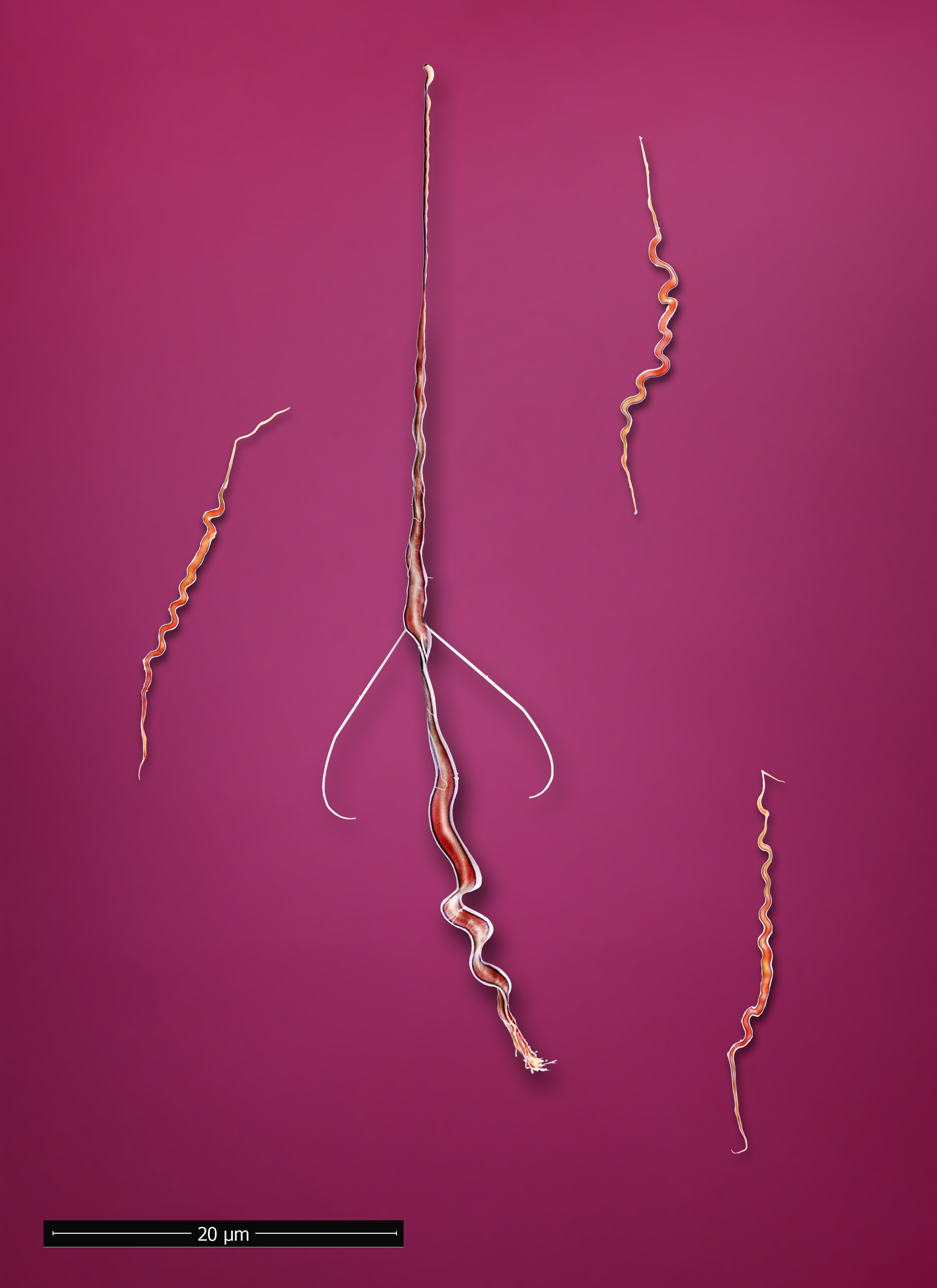
Here we see a cut through the same animal but more posterior. On the top there is an area where cilia from the epidermis are moving inwards. That is the male genital opening and the large U-shaped structure behind it is the male stylet (copulatory organ). To the right of the stylet is the seminal vesicle, where sperm is stored before copulation. As you can see Macrostomum sperm is quite large. In some species the sperm can be almost as long as the stylet.
Since the serial sections allow us to trace structures along the animal we can also reconstruct them. Here you can see a 3D reconstruction of the male copulatory organ - the stylet. It has quite a complex structure with a complicate slightly corkscrew shaped curve to it. From 2D images it is difficult to completely grasp the subtleties of this turn but we can see it quite easily in the reconstruction. In 3D we see this quite clearly. Here is an example from a species we discovered in Lake Tanganyika, Zambia on a recent field trip:
{% include youtubePlayer.html id="ZE5gi63MnPU" %}
Together with Martin Oeggerli we have made some SEM images of sperm and Martin them arranged an colored them:


